
Prostate Scan
What is a Prostate Scan?
A prostate scan is a diagnostic imaging test that assesses the size, shape, structure, and function of the prostate gland. The prostate is a small gland in men located below the bladder and plays a role in producing seminal fluid. As men age, the prostate can become enlarged or develop conditions such as inflammation, benign growth, or cancer.
A prostate scan allows clinicians to detect abnormalities early, guide diagnosis, and plan appropriate treatment. It is commonly used in men who have urinary symptoms, elevated prostate-specific antigen (PSA) levels, or concerns about prostate cancer.
Who is Suitable for a Prostate Scan?
A prostate scan is recommended for men who may be at risk of prostate conditions or who are experiencing symptoms suggestive of prostate disease.
Common indications include:
- Men with urinary symptoms: Difficulty urinating, weak urine flow, frequent urination (especially at night), or incomplete bladder emptying.
- Elevated PSA levels: A blood test showing increased prostate-specific antigen may indicate inflammation, enlargement, or cancer.
- Abnormal digital rectal examination (DRE): If a doctor detects irregularities during a physical exam.
- Family history of prostate cancer: Particularly if close relatives have been affected.
- Monitoring known prostate conditions: Such as benign prostatic hyperplasia (BPH) or previously diagnosed prostate cancer.
- Post-treatment follow-up: To assess response after surgery, radiation, or other therapies.
Men over the age of 50 are more commonly assessed, although younger men with symptoms or risk factors may also require evaluation.
Benefits of a Prostate Scan
A prostate scan offers several important clinical and patient-focused advantages, particularly in early detection and accurate diagnosis.
- Early Detection of Prostate Cancer: Helps identify cancer at an early stage when treatment is more effective.
- Improved Diagnostic Accuracy: Advanced imaging, especially MRI, can distinguish between benign and suspicious areas.
- Guidance for Biopsy: Scans can pinpoint specific areas of concern, improving the accuracy of targeted biopsies.
- Non-Invasive Assessment: Most prostate scans do not require surgery or invasive procedures.
- Treatment Planning: Helps determine the size, location, and extent of disease, guiding personalised treatment decisions.
- Monitoring Disease Progression: Useful for tracking changes over time, particularly in active surveillance strategies.
- Reduced Unnecessary Procedures: High-quality imaging can sometimes avoid unnecessary biopsies in low-risk patients.
From a patient perspective, prostate scans provide reassurance, clarity, and a structured pathway for diagnosis and management.
Types of Prostate Scan
There are several types of prostate scans, each with specific uses depending on the clinical situation.
Multiparametric MRI (mpMRI) of the Prostate
- Most advanced and commonly used imaging tool for prostate cancer detection
- Combines different MRI techniques to assess tissue structure, blood flow, and cellular activity
- Helps identify suspicious lesions and assess cancer aggressiveness
- Widely used before biopsy and for active surveillance
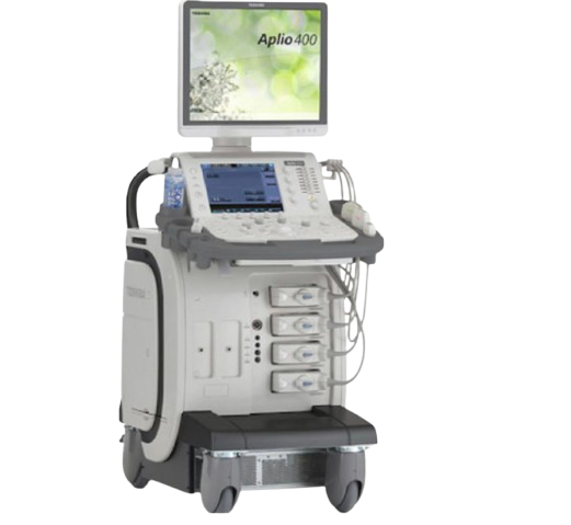
Transrectal Ultrasound (TRUS)
- Uses sound waves via a probe inserted into the rectum
- Commonly used to guide prostate biopsies
- Useful for measuring prostate size and detecting enlargement
- Less sensitive than MRI for detecting cancer, but widely accessible
Abdominal or Pelvic Ultrasound
- Non-invasive scan performed over the lower abdomen
- Helps assess bladder function and prostate enlargement
- Often used as an initial screening tool
Computed Tomography (CT Scan)
- Not typically used for primary prostate diagnosis
- Useful in assessing the spread of cancer to lymph nodes or other organs
- May be part of staging in advanced disease
PSMA PET Scan (Prostate-Specific Membrane Antigen PET)
- Highly specialised nuclear medicine scan
- Detects prostate cancer cells with high sensitivity
- Often used for staging or detecting recurrence
- Particularly valuable in advanced or high-risk cases
Bone Scan
- Used when there is concern that prostate cancer may have spread to the bones
- Helps detect metastases and guide treatment planning
Preparation for a Prostate Scan
Preparation for a prostate scan depends on the type of imaging being performed. Most prostate scans are straightforward and require minimal preparation, but following instructions carefully helps ensure accurate results and a smooth experience.
- Confirm the Type of Scan: Your doctor or imaging provider will advise whether you are having an MRI, ultrasound (TRUS), CT, or a specialised scan such as PSMA PET. Preparation varies slightly for each.
- Provide Medical History: Inform your doctor about any previous prostate conditions, surgeries, allergies (especially to contrast dye), and current medications. This is particularly important for scans involving contrast agents.
- PSA and Blood Tests: In many cases, your scan is arranged after a prostate-specific antigen (PSA) test. Ensure your results are available for comparison.
- Medication Instructions:
- Regular medications: Usually continued unless advised otherwise
- Blood thinners: May need adjustment if a biopsy is planned alongside imaging
- Diabetes medications: May need modification if fasting is required
- Fasting (if required): Some scans, particularly MRI with contrast or PET scans, may require fasting for a few hours before the procedure.
- Bowel Preparation (for TRUS or MRI): You may be asked to use a mild enema to clear the rectum for better image quality.
- Bladder Preparation:
- Some scans require a comfortably full bladder
- Others may require you to empty your bladder beforehand
- Remove Metal Objects: For MRI scans, remove jewellery, watches, belts, and any metal-containing items. Notify staff if you have implants, pacemakers, or metal fragments.
- Clothing: Wear loose, comfortable clothing, or you may be asked to change into a gown.
- Address Anxiety or Claustrophobia: If you feel anxious about enclosed spaces (MRI), discuss this beforehand—sedation options may be available.
Preparation is generally simple, and most patients can continue normal activities before the scan unless specifically instructed otherwise.
Prostate Scan Procedure
The procedure varies depending on the type of scan, but most prostate scans are painless and performed on an outpatient basis.
During a Prostate MRI
- You lie on a scanning table that slides into the MRI machine
- A coil (receiver device) may be placed around your pelvic area
- The scan takes 20–45 minutes
- You must remain still while images are taken
- Loud tapping or buzzing noises are normal (ear protection is provided)
- A contrast injection may be given through a vein to enhance imaging
During a Transrectal Ultrasound (TRUS)
- A small lubricated probe is gently inserted into the rectum
- The scan takes about 10–20 minutes
- You may feel pressure, but it is usually not painful
- Often used to guide prostate biopsy if needed
During a CT Scan
- You lie on a table that moves through a circular scanner
- The scan is quick (usually under 10 minutes)
- Contrast dye may be injected to improve image clarity
During a PSMA PET Scan
- A small radioactive tracer is injected into your bloodstream
- You wait for the tracer to circulate (30–60 minutes)
- The scan itself takes 20–40 minutes
- Images show areas of increased prostate cancer activity
What to Expect After a Prostate Scan?
Recovery after a prostate scan is typically immediate, with most patients resuming normal activities the same day.
- Return to Normal Activities: You can usually drive, work, and eat as normal unless sedation was used.
- Hydration: If contrast dye was used, drinking fluids helps flush it from your system.
- Mild Discomfort (TRUS only):
- Temporary rectal discomfort or pressure
- Minor spotting if a biopsy was performed
- No Radiation Effects (MRI/Ultrasound): These scans do not use ionising radiation.
- Radiation Precautions (PET/CT): Minimal exposure; generally safe with no special precautions required
- Results Timeline: Results are usually available within 24–72 hours. Your doctor will review findings and discuss next steps
- Follow-Up Plan:
- May include further imaging, biopsy, or monitoring
- Results are often combined with PSA levels and clinical findings
Prostate Scan Prognosis
A prostate scan itself does not determine prognosis but plays a critical role in shaping it by enabling early and accurate diagnosis.
- Early Detection Improves Outcomes: Identifying prostate cancer early often leads to highly effective treatment and long-term survival.
- Risk Stratification: Advanced imaging helps classify disease as low, intermediate, or high risk, guiding treatment decisions.
- Active Surveillance: For low-risk cases, scans support safe monitoring without immediate intervention.
- Treatment Planning: Imaging defines tumour location and spread, helping tailor surgery, radiation, or medical therapy.
- Monitoring Response: Repeated scans can track treatment effectiveness over time.
In modern clinical practice, particularly with multiparametric MRI and PSMA PET scans, prognosis has improved due to more precise and personalised care pathways.
Prostate Scan Risks
Prostate scans are generally safe, but, like all medical procedures, they carry some potential risks depending on the type of scan.
- MRI Risks:
- Rare allergic reaction to contrast dye
- Issues with metal implants ,if not disclosed
- Claustrophobia or discomfort in enclosed spaces
- Ultrasound (TRUS) Risks:
- Mild discomfort during probe insertion
- Small risk of infection if biopsy is performed
- Minor bleeding (temporary)
- CT Scan Risks:
- Exposure to ionising radiation (low dose)
- Possible reaction to contrast dye
- PSMA PET Scan Risks:
- Low-level radiation exposure
- Rare allergic reaction to tracer
- General Considerations:
- Most risks are minimal and temporary
- Benefits of accurate diagnosis typically outweigh risks
What if a Prostate Scan is Delayed?
Delaying a prostate scan can lead to missed or late diagnosis of conditions such as prostate cancer or significant prostate enlargement, which may progress silently without obvious symptoms. This delay can reduce the opportunity for early, less invasive treatment and may result in more complex management if the condition advances. Urinary symptoms may worsen over time, and patients may experience increased anxiety due to uncertainty.
While some prostate conditions are slow-growing, timely imaging is important to ensure accurate assessment, guide appropriate care, and maintain the best possible long-term outcomes.