
Doppler Vascular Ultrasound
What is Doppler Vascular Ultrasound?
Doppler Vascular Ultrasound is a non-invasive imaging test that uses high-frequency sound waves to assess blood flow through your arteries and veins. It is an advanced form of standard ultrasound that incorporates the Doppler effect, allowing clinicians to measure the speed and direction of blood flow in real time.
Unlike a conventional ultrasound, which primarily shows tissue and organ structure, a Doppler ultrasound provides functional information. It can detect how well blood is moving through vessels, whether there are blockages, narrowing (stenosis), or abnormal flow patterns.
This test is commonly used to examine blood vessels in areas such as the neck (carotid arteries), arms, legs, abdomen, and, sometimes, organs such as the kidneys. It plays an essential role in diagnosing vascular conditions and guiding treatment decisions.
Doppler Vascular Ultrasound is safe, painless, and radiation-free, making it suitable for repeated use and a wide range of patients.
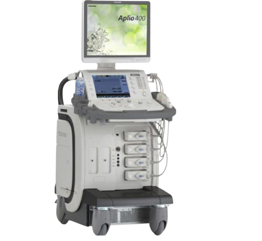
Who is Suitable for Doppler Vascular Ultrasound?
Doppler Vascular Ultrasound is recommended for individuals with symptoms or risk factors for poor circulation or vascular disease.
- Patients with leg pain or swelling: May indicate conditions such as deep vein thrombosis (DVT) or venous insufficiency.
- Individuals with suspected arterial disease: Symptoms like leg pain when walking (claudication) can suggest peripheral artery disease.
- People with stroke risk factors: Used to assess carotid arteries in patients with high blood pressure, diabetes, or prior stroke/TIA.
- Patients with varicose veins: Helps evaluate vein function and valve competence.
- Individuals with unexplained limb coldness or colour changes: May indicate reduced blood flow.
- Patients undergoing vascular surgery planning or follow-up: Used before and after procedures such as bypass surgery or stenting.
- People with abdominal or renal concerns: Can assess blood flow to organs like the kidneys.
- Pregnant patients (in specific cases): Sometimes used to evaluate blood flow in certain clinical situations.
It is also suitable for screening in high-risk individuals, particularly those with a history of smoking, high cholesterol, or cardiovascular disease.
Benefits of Doppler Vascular Ultrasound
Doppler Vascular Ultrasound offers several important clinical and patient-focused advantages.
- Non-invasive and painless: No needles, incisions, or injections are typically required.
- No radiation exposure: Unlike CT scans, it is safe for repeated use and for vulnerable populations.
- Real-time blood flow assessment: Provides immediate insight into how blood is moving through vessels.
- Early detection of vascular disease: Can identify narrowing, blockages, or clots before symptoms become severe.
- Guides treatment decisions: Helps determine whether medical management, lifestyle changes, or procedures are needed.
- Portable and accessible: Often performed in clinics, hospitals, or even bedside settings.
- Cost-effective: Generally less expensive than advanced imaging such as CT or MRI.
- Safe for most patients: Including those with kidney issues or contrast allergies.
From a clinical perspective, Doppler ultrasound is often the first-line investigation for many vascular conditions due to its safety and effectiveness.
Types of Doppler Vascular Ultrasound
There are several types of Doppler ultrasound techniques, each providing different types of information about blood flow.
- Colour Doppler Ultrasound: Displays blood flow direction and velocity using colours (commonly red and blue). It helps visualise flow patterns within vessels and detect abnormalities.
- Spectral Doppler (Pulse Wave Doppler): Provides a graphical representation (waveform) of blood flow velocity over time. This is useful for measuring the severity of narrowing or blockages.
- Duplex Ultrasound: Combines traditional ultrasound imaging with Doppler flow analysis. It allows clinicians to see both the structure of the vessel and the blood flow within it simultaneously.
- Continuous Wave Doppler: Measures high-velocity blood flow and is often used in specialised vascular or cardiac assessments.
- Power Doppler: More sensitive in detecting low-flow blood, useful in small vessels or organs where flow may be subtle.
Alternative Options to Doppler Vascular Ultrasound
While Doppler Vascular Ultrasound is often the first-line investigation, there are alternative imaging modalities that may be used depending on the clinical situation.
- CT Angiography (CTA)
- Magnetic Resonance Angiography (MRA)
- Conventional Angiography
- Standard Ultrasound (Non-Doppler)
- Ankle-Brachial Index (ABI)
- Nuclear Medicine Scans
Each alternative has its advantages and limitations. In most cases, Doppler Vascular Ultrasound is used first due to its safety and accessibility, with other tests reserved for more complex or unclear cases.
Preparation for a Doppler Vascular Ultrasound
Preparation for a Doppler Vascular Ultrasound is usually simple and depends on the area of the body being examined. In most cases, minimal preparation is required, making it a convenient and accessible investigation.
- Follow specific instructions if provided: Some scans, such as abdominal vascular studies, may require fasting for several hours to improve image quality.
- Wear comfortable, loose clothing: This allows easy access to the area being examined, such as the neck, arms, or legs.
- Avoid smoking before the test: Nicotine can temporarily affect blood flow and may influence results, particularly in vascular studies.
- Continue regular medications unless advised otherwise: Most patients can take their usual medications without interruption.
- Stay well hydrated: Good hydration can sometimes assist with clearer imaging of blood vessels.
- Bring relevant medical information: Previous imaging, referral letters, and a list of medications may help guide the examination.
For most vascular Doppler studies (e.g., legs or carotid arteries), no fasting or special preparation is needed. The radiology team will provide tailored instructions if your scan requires any additional steps.
Doppler Vascular Ultrasound Procedure
A Doppler Vascular Ultrasound is a straightforward and painless procedure performed by a trained sonographer or radiologist.
- Positioning: You will be asked to lie on an examination bed or sit comfortably, depending on the area being scanned.
- Application of gel: A clear, water-based gel is applied to the skin. This helps transmit sound waves and ensures accurate imaging.
- Use of transducer (probe): A handheld device is gently moved over the skin. It emits sound waves that reflect off blood cells and vessel walls.
- Real-time imaging and sound: The machine displays images and may produce a “whooshing” sound, representing blood flow through the vessels.
- Assessment of blood flow: The clinician may apply light pressure or adjust your position to evaluate flow from different angles.
- Breathing or movement instructions: You may be asked to hold your breath or change position briefly to improve image quality.
The procedure typically takes 20 to 45 minutes, depending on the complexity and number of vessels being assessed. There is no pain, although slight pressure from the probe may be felt.
What to Expect After a Doppler Vascular Ultrasound?
After the scan, most patients can return to their normal daily activities immediately without restrictions.
- No recovery time required: The procedure is non-invasive, so you can resume work, driving, and usual routines straight away.
- Gel removal: The ultrasound gel is wiped off your skin before you leave.
- Immediate comfort: There are no lingering effects such as pain or sedation.
- Results processing: Images are reviewed by a radiologist, and a report is sent to your referring doctor.
- Follow-up plan: Your doctor will discuss the findings and recommend next steps, which may include monitoring, lifestyle changes, or further investigations.
In urgent cases, preliminary findings may be communicated quickly, especially if there is concern about conditions such as blood clots.
Doppler Vascular Ultrasound Prognosis
The prognosis associated with Doppler Vascular Ultrasound is not about the test itself, but rather how effectively it contributes to diagnosis and management.
- Early detection improves outcomes: Identifying conditions like arterial narrowing or venous clots early allows timely treatment and reduces complications.
- Guides appropriate treatment: Results help determine whether conservative management, medication, or intervention is required.
- Monitors disease progression: Repeat scans can track changes in blood flow over time, helping assess treatment effectiveness.
- Supports preventive care: Detecting vascular issues early can prompt lifestyle modifications, reducing long-term cardiovascular risk.
Doppler ultrasound plays a critical role in improving patient outcomes by enabling accurate, early, and ongoing assessment of vascular health.
Doppler Vascular Ultrasound Risks
Doppler Vascular Ultrasound is considered extremely safe, with very few associated risks.
- No radiation exposure: Unlike CT scans, it does not use ionising radiation.
- Non-invasive: No needles or surgical procedures are involved.
- Minimal discomfort: Some mild pressure from the probe may be felt, particularly over sensitive areas.
- Rare limitations rather than risks: Factors such as obesity, swelling, or deep vessels may affect image clarity but do not pose harm.
- No known long-term side effects: Ultrasound technology has been widely used for decades with an excellent safety record.
Because of its safety profile, Doppler ultrasound is often the preferred first-line investigation for vascular conditions.
What if Doppler Vascular Ultrasound is Delayed?
Delaying a Doppler Vascular Ultrasound can have varying consequences depending on the underlying condition being investigated.
- Delayed diagnosis of serious conditions
- Increased risk of complications
- Progression of disease
- Persistent symptoms
- Missed opportunity for early intervention
In situations where symptoms are acute—such as sudden leg swelling, chest pain, or neurological symptoms—prompt imaging is important and should not be delayed.