
Breast Radiology
What is Breast Radiology?
Breast Radiology is a specialised area of medical imaging focused on assessing the structure and health of breast tissue. It plays a critical role in the early detection, diagnosis, and monitoring of breast conditions, particularly Breast Cancer.
This field combines advanced imaging technologies with expert interpretation by trained radiologists to identify abnormalities such as lumps, cysts, calcifications, or structural changes that may not be visible or palpable during a physical examination.
Breast Radiology is most commonly used for:
- Screening: Detecting early signs of disease before symptoms develop
- Diagnosis: Investigating symptoms such as lumps, pain, or nipple discharge
- Monitoring: Tracking known conditions over time
- Guidance: Assisting in procedures such as biopsies
The most widely recognised imaging modality within Breast Radiology is Mammography, but it also includes ultrasound, MRI, and image-guided interventions.
Importantly, Breast Radiology is not limited to cancer detection. It also evaluates benign (non-cancerous) conditions such as fibrocystic changes, infections, and hormonal-related changes in the breast.
Who is Suitable for Breast Radiology?
Breast Radiology is suitable for a broad range of individuals, both for routine screening and for diagnostic purposes.
- Women aged 40 and above: Regular screening is recommended in many countries to detect early-stage breast cancer, often before symptoms appear.
- Women with a family history of breast cancer: Those with genetic predispositions (such as BRCA mutations) may require earlier and more frequent imaging.
- Patients with breast symptoms: This includes individuals experiencing:
- Lumps or thickening
- Breast pain
- Skin changes or dimpling
- Nipple discharge or inversion
- Patients with dense breast tissue: Dense tissue can make abnormalities harder to detect on mammograms, so additional imaging, such as ultrasound or MRI, may be advised.
- Patients undergoing treatment or follow-up: Breast Radiology is used to monitor treatment response and detect recurrence.
- Men with breast symptoms: Although less common, men can develop breast conditions, including cancer and may require imaging assessment.
- Patients requiring image-guided procedures: Radiology is essential for accurate guidance of biopsies or aspirations.
Suitability is typically determined by a referring doctor based on individual risk factors, symptoms, and clinical findings.
Benefits of Breast Radiology
Breast Radiology provides several important clinical and patient-centred benefits.
- Early Detection of Disease: Breast imaging can identify abnormalities before they become clinically apparent. Early detection of Breast Cancer significantly improves treatment outcomes and survival rates.
- Non-Invasive Assessment: Most breast imaging techniques are non-invasive and do not require surgery, making them safe and accessible for routine screening.
- Accurate Diagnosis: Combining different imaging modalities allows for a more precise evaluation of breast tissue, helping distinguish between benign and malignant conditions.
- Guidance for Procedures: Imaging can guide minimally invasive procedures such as biopsies, ensuring accurate sampling of suspicious areas.
- Monitoring Over Time: Breast Radiology enables clinicians to track changes in breast tissue, evaluate treatment effectiveness, and detect recurrence early.
- Personalised Care Planning: Imaging findings help tailor treatment strategies based on the individual’s condition, risk profile, and disease progression.
- Improved Patient Reassurance: For many patients, imaging provides peace of mind when symptoms are investigated and found to be benign.
Types of Breast Radiology
There are several types of imaging used in Breast Radiology, each suited to different clinical needs.
- Mammography: A low-dose X-ray of the breast and the most common screening tool. It is highly effective in detecting early breast cancer, particularly in women over 40.
- Breast Ultrasound: Uses sound waves to create images. It is often used to assess lumps detected on mammography or physical examination and is particularly useful in younger women with denser breast tissue.
- Breast MRI: Uses magnetic fields and contrast dye to produce highly detailed images. It is typically used for high-risk patients or when further clarification is needed after other imaging.
- Digital Breast Tomosynthesis (3D Mammography): Provides a three-dimensional view of the breast, improving detection rates and reducing false positives.
- Image-Guided Procedures: Includes biopsies performed under ultrasound, mammography, or MRI guidance to obtain tissue samples for analysis.
Each imaging modality has a specific role, and often they are used together to provide a comprehensive evaluation.
Alternative Options to Breast Radiology
While Breast Radiology is the gold standard for breast imaging, complementary or alternative approaches may be appropriate in certain situations.
- Clinical breast examination: Physical assessment by a healthcare provider
- Self-breast awareness: Monitoring for changes in size, shape, or feel
- Genetic testing: For individuals with a strong family history
- Blood-based biomarkers (emerging): Currently under research but not widely used
However, these methods do not replace imaging and are typically used alongside Breast Radiology rather than instead of it.
Preparation for Breast Radiology
Preparation depends on the type of imaging being performed, but general recommendations include:
- Schedule appropriately: Mammograms are often best performed one week after your menstrual period, when breasts are less tender
- Avoid deodorants or powders: These can interfere with imaging results, particularly for mammography
- Inform your provider: About pregnancy, breastfeeding, or implants
- Bring previous imaging: For comparison, if available
- Wear comfortable clothing: You may need to undress from the waist up
Clear communication with your healthcare provider ensures the most accurate and safe imaging experience.
Breast Radiology Procedure
The experience varies depending on the type of imaging performed, but most procedures are quick and well-tolerated.
Mammography
During Mammography:
- Each breast is placed on a flat plate and gently compressed
- Compression lasts only a few seconds and improves image clarity
- Images are taken from multiple angles
- The procedure typically takes 10–20 minutes
You may feel pressure or mild discomfort, but it is brief and necessary for accurate results.
Breast Ultrasound
During Breast Ultrasound:
- You will lie on an examination bed
- A gel is applied to the skin
- A handheld probe is moved over the breast area
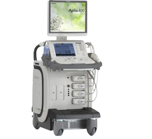
- Real-time images are displayed on a screen
This test is painless and usually takes 15–30 minutes.
Breast MRI
During Breast MRI:
- You lie face down on a special table with openings for the breasts
- The table slides into the MRI scanner
- A contrast dye may be injected through an IV line
- You must remain still while images are taken
The scan takes approximately 30–45 minutes. Some patients may feel confined or hear loud noises, but ear protection is provided.
Image-Guided Biopsy (if required)
If an abnormality is detected:
- Local anaesthetic is applied
- Imaging guides a needle to the target area
- A small tissue sample is collected
This is a minimally invasive procedure and is typically completed within an hour.
What to Expect After Breast Imaging?
Recovery and follow-up depend on the type of imaging performed.
Immediate recovery
- Most patients can resume normal activities straight away
- No downtime is required after mammography, ultrasound, or MRI
- Mild tenderness may occur after compression during mammography
After a biopsy
- Slight bruising, swelling, or discomfort may occur
- A small dressing is applied
- Strenuous activity may be limited for 24–48 hours
Receiving results
- Some results may be available on the same day
- Others are reviewed by a radiologist and sent to your referring doctor
- Follow-up appointments may be arranged if needed
Next steps (if abnormalities are found)
- Additional imaging
- Short-term monitoring
- Biopsy or specialist referral
Many findings are benign, and further testing is often precautionary.
Breast Radiology Prognosis
Breast Radiology significantly improves health outcomes by enabling early detection and accurate diagnosis.
- Early-stage detection: Leads to higher survival rates and less aggressive treatment
- Better monitoring: Supports long-term management of breast conditions
- Personalised care: Imaging results guide tailored treatment plans
When used as part of regular screening, Breast Radiology plays a vital role in preventative healthcare and ongoing monitoring.
Breast Radiology Risks
Breast Radiology is generally very safe, but there are some minimal risks to consider.
- Low radiation exposure: From mammography, although levels are considered safe
- False positives: May lead to additional testing or anxiety
- False negatives: Rarely, abnormalities may not be detected
- Contrast reactions: In MRI, allergic reactions to contrast dye are uncommon
- Discomfort: Temporary discomfort during compression or procedures
The benefits of early detection typically outweigh these small risks.
What if Breast Radiology is Delayed?
Delaying Breast Radiology can lead to missed or late diagnosis of potentially serious conditions, including breast cancer. Without timely imaging, abnormalities may go undetected and progress to more advanced stages, limiting treatment options and reducing the likelihood of successful outcomes.
Early detection is one of the most important factors in improving survival rates, so postponing recommended imaging may result in more complex treatments, increased health risks, and poorer long-term prognosis.